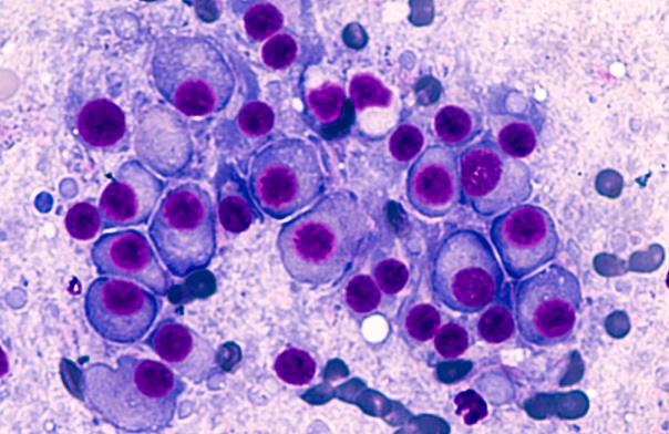
Histopathology is the study of change in diseased tissues using a microscopic examination in a biopsy or a surgical specimen. That tissue will be processed and put into a glass slide before inspection. Nuclei segmentation in histopathology images is a very crucial step in cancer diagnosis and grading since it can be a powerful tool in providing the proper treatment.
However, there are certain challenges to histopathological images such as:
- Whole slide images (WSI) are very large in size (ranges between 20,000x20,000 pixels to over 100,000x100,000 pixels)
- Insufficient annotated images
- Dealing with overlapping nuclei
- Diversity of Microscopic images
The aim of this project is to improve on nuclei segmentation methods by tackling the problems previously mentioned, which will eventually lead to computer-diagnosis systems that identify and grade cancer.
Team Members